刺激
Stimulus
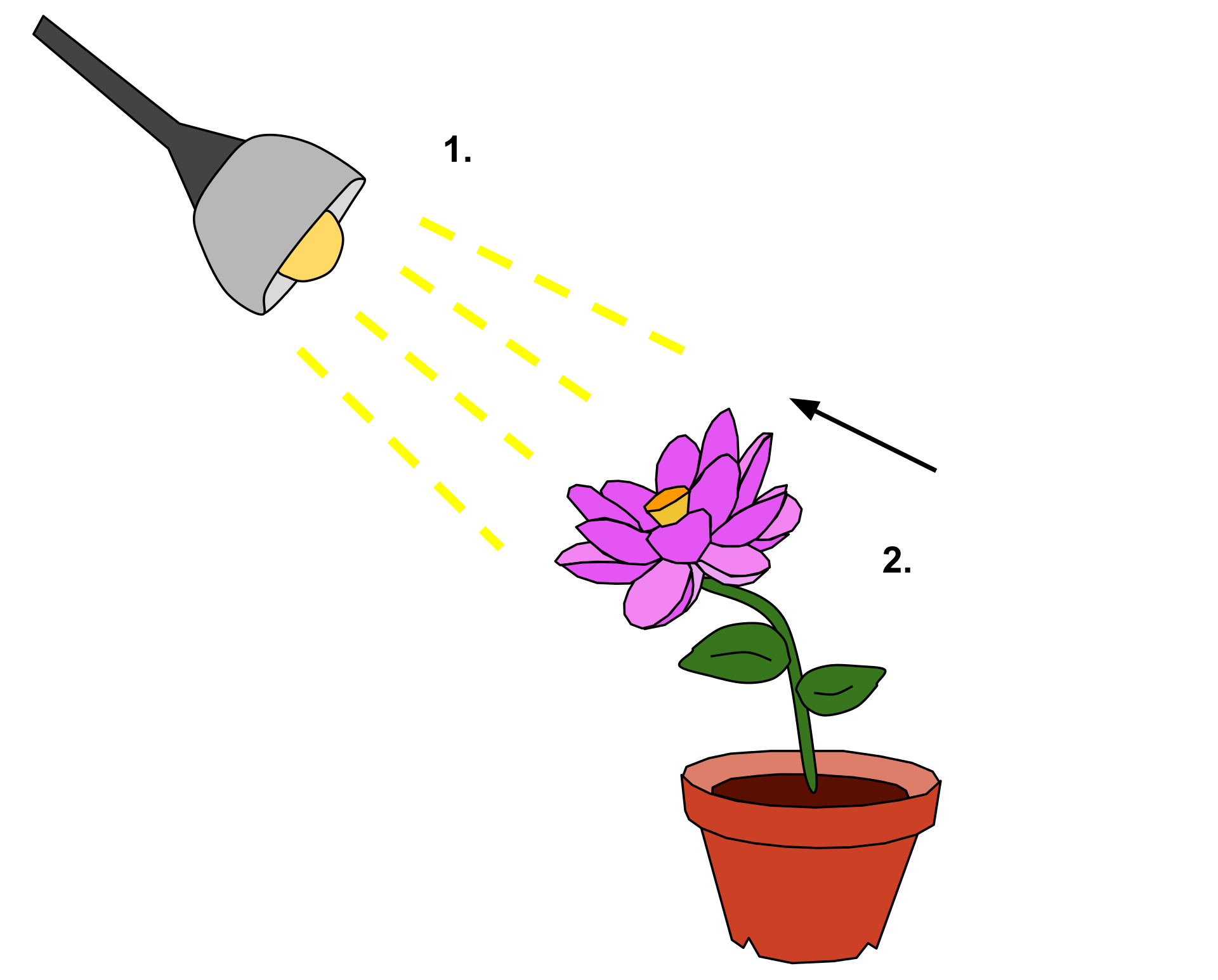
The
light from the lamp (1.) functions as a detectable change in the
plant's environment. As a result, the plant exhibits a reaction of
phototropism—directional growth (2.) toward the light stimulus.
☆ 生理学において刺激(stimulus)とは、生物の内部または外部環境の物理的または化学的構造における検出可能な変化のことである。感覚受容体は、皮膚にある触覚受 容体や眼球にある光受容体のように体外から情報を受け取る場合と、化学受容体や機械受容体のように体内から情報を受け取る場合がある。刺激が感覚受容器に よって検出されると、刺激伝達を 介して反射を引き起こすことができる。内部刺激は多くの場合、恒常性制御システムの最初の構成要素である。外部からの刺激は、闘争・逃走反応のように、全 身的な 反応を全身に引き起こすことができる。刺激が高い確率で検出されるためには、その強さのレベルが絶対的閾値を超えなければならない。信号が閾値に達した場 合、その情報は中枢神経系(CNS)に伝達され、そこで統合され、どのように反応するかの決定がなされる。刺激は一般的に身体を反応させるが、信号が反応 を引き起こすかどうかを最終的に決定するのはCNSである。
| In physiology, a stimulus[1]
is a detectable change in the physical or chemical structure of an
organism's internal or external environment. The ability of an organism
or organ to detect external stimuli, so that an appropriate reaction
can be made, is called sensitivity (excitability).[2] Sensory receptors
can receive information from outside the body, as in touch receptors
found in the skin or light receptors in the eye, as well as from inside
the body, as in chemoreceptors and mechanoreceptors. When a stimulus is
detected by a sensory receptor, it can elicit a reflex via stimulus
transduction. An internal stimulus is often the first component of a
homeostatic control system. External stimuli are capable of producing
systemic responses throughout the body, as in the fight-or-flight
response. In order for a stimulus to be detected with high probability,
its level of strength must exceed the absolute threshold; if a signal
does reach threshold, the information is transmitted to the central
nervous system (CNS), where it is integrated and a decision on how to
react is made. Although stimuli commonly cause the body to respond, it
is the CNS that finally determines whether a signal causes a reaction
or not. |
生理学において刺激
[1]とは、生物の内部または外部環境の物理的または化学的構造における検出可能な変化のことである。感覚受容体は、皮膚にある触覚受容体や眼球にある光
受容体のように体外から情報を受け取る場合と、化学受容体や機械受容体のように体内から情報を受け取る場合がある。刺激が感覚受容器によって検出される
と、刺激伝達を
介して反射を引き起こすことができる。内部刺激は多くの場合、恒常性制御システムの最初の構成要素である。外部からの刺激は、闘争・逃走反応のように、全
身的な
反応を全身に引き起こすことができる。刺激が高い確率で検出されるためには、その強さのレベルが絶対的閾値を超えなければならない。信号が閾値に達した場
合、その情報は中枢神経系(CNS)に伝達され、そこで統合され、どのように反応するかの決定がなされる。刺激は一般的に身体を反応させるが、信号が反応
を引き起こすかどうかを最終的に決定するのはCNSである。 |
| Types Internal Homeostatic imbalances Homeostatic outbalances are the main driving force for changes of the body. These stimuli are monitored closely by receptors and sensors in different parts of the body. These sensors are mechanoreceptors, chemoreceptors and thermoreceptors that, respectively, respond to pressure or stretching, chemical changes, or temperature changes. Examples of mechanoreceptors include baroreceptors which detect changes in blood pressure, Merkel's discs which can detect sustained touch and pressure, and hair cells which detect sound stimuli. Homeostatic imbalances that can serve as internal stimuli include nutrient and ion levels in the blood, oxygen levels, and water levels. Deviations from the homeostatic ideal may generate a homeostatic emotion, such as pain, thirst or fatigue, that motivates behavior that will restore the body to stasis (such as withdrawal, drinking or resting).[3] Blood pressure Blood pressure, heart rate, and cardiac output are measured by stretch receptors found in the carotid arteries. Nerves embed themselves within these receptors and when they detect stretching, they are stimulated and fire action potentials to the central nervous system. These impulses inhibit the constriction of blood vessels and lower the heart rate. If these nerves do not detect stretching, the body determines perceives low blood pressure as a dangerous stimulus and signals are not sent, preventing the inhibition CNS action; blood vessels constrict and the heart rate increases, causing an increase in blood pressure in the body.[4] External Touch and pain Sensory feelings, especially pain, are stimuli that can elicit a large response and cause neurological changes in the body. Pain also causes a behavioral change in the body, which is proportional to the intensity of the pain. The feeling is recorded by sensory receptors on the skin and travels to the central nervous system, where it is integrated and a decision on how to respond is made; if it is decided that a response must be made, a signal is sent back down to a muscle, which behaves appropriately according to the stimulus.[3] The postcentral gyrus is the location of the primary somatosensory area, the main sensory receptive area for the sense of touch.[5] Pain receptors are known as nociceptors. Two main types of nociceptors exist, A-fiber nociceptors and C-fiber nociceptors. A-fiber receptors are myelinated and conduct currents rapidly. They are mainly used to conduct fast and sharp types of pain. Conversely, C-fiber receptors are unmyelinated and slowly transmit. These receptors conduct slow, burning, diffuse pain.[6] The absolute threshold for touch is the minimum amount of sensation needed to elicit a response from touch receptors. This amount of sensation has a definable value and is often considered to be the force exerted by dropping the wing of a bee onto a person's cheek from a distance of one centimeter. This value will change based on the body part being touched.[7] Vision Vision provides opportunity for the brain to perceive and respond to changes occurring around the body. Information, or stimuli, in the form of light enters the retina, where it excites a special type of neuron called a photoreceptor cell. A local graded potential begins in the photoreceptor, where it excites the cell enough for the impulse to be passed along through a track of neurons to the central nervous system. As the signal travels from photoreceptors to larger neurons, action potentials must be created for the signal to have enough strength to reach the CNS.[4] If the stimulus does not warrant a strong enough response, it is said to not reach absolute threshold, and the body does not react. However, if the stimulus is strong enough to create an action potential in neurons away from the photoreceptor, the body will integrate the information and react appropriately. Visual information is processed in the occipital lobe of the CNS, specifically in the primary visual cortex.[4] The absolute threshold for vision is the minimum amount of sensation needed to elicit a response from photoreceptors in the eye. This amount of sensation has a definable value and is often considered to be the amount of light present from someone holding up a single candle 30 miles away, if one's eyes were adjusted to the dark.[7] Smell Smell allows the body to recognize chemical molecules in the air through inhalation. Olfactory organs located on either side of the nasal septum consist of olfactory epithelium and lamina propria. The olfactory epithelium, which contains olfactory receptor cells, covers the inferior surface of the cribiform plate, the superior portion of the perpendicular plate, the superior nasal concha. Only roughly two percent of airborne compounds inhaled are carried to olfactory organs as a small sample of the air being inhaled. Olfactory receptors extend past the epithelial surface providing a base for many cilia that lie in the surrounding mucus. Odorant-binding proteins interact with these cilia stimulating the receptors. Odorants are generally small organic molecules. Greater water and lipid solubility is related directly to stronger smelling odorants. Odorant binding to G protein coupled receptors activates adenylate cyclase, which converts ATP to camp. cAMP, in turn, promotes the opening of sodium channels resulting in a localized potential.[8] The absolute threshold for smell is the minimum amount of sensation needed to elicit a response from receptors in the nose. This amount of sensation has a definable value and is often considered to be a single drop of perfume in a six-room house. This value will change depending on what substance is being smelled.[7] Taste Taste records flavoring of food and other materials that pass across the tongue and through the mouth. Gustatory cells are located on the surface of the tongue and adjacent portions of the pharynx and larynx. Gustatory cells form on taste buds, specialized epithelial cells, and are generally turned over every ten days. From each cell, protrudes microvilli, sometimes called taste hairs, through also the taste pore and into the oral cavity. Dissolved chemicals interact with these receptor cells; different tastes bind to specific receptors. Salt and sour receptors are chemically gated ion channels, which depolarize the cell. Sweet, bitter, and umami receptors are called gustducins, specialized G protein coupled receptors. Both divisions of receptor cells release neurotransmitters to afferent fibers causing action potential firing.[8] The absolute threshold for taste is the minimum amount of sensation needed to elicit a response from receptors in the mouth. This amount of sensation has a definable value and is often considered to be a single drop of quinine sulfate in 250 gallons of water.[7] Sound Changes in pressure caused by sound reaching the external ear resonate in the tympanic membrane, which articulates with the auditory ossicles, or the bones of the middle ear. These tiny bones multiply these pressure fluctuations as they pass the disturbance into the cochlea, a spiral-shaped bony structure within the inner ear. Hair cells in the cochlear duct, specifically the organ of Corti, are deflected as waves of fluid and membrane motion travel through the chambers of the cochlea. Bipolar sensory neurons located in the center of the cochlea monitor the information from these receptor cells and pass it on to the brainstem via the cochlear branch of cranial nerve VIII. Sound information is processed in the temporal lobe of the CNS, specifically in the primary auditory cortex.[8] The absolute threshold for sound is the minimum amount of sensation needed to elicit a response from receptors in the ears. This amount of sensation has a definable value and is often considered to be a watch ticking in an otherwise soundless environment 20 feet away.[7] Equilibrium Semi circular ducts, which are connected directly to the cochlea, can interpret and convey to the brain information about equilibrium by a similar method as the one used for hearing. Hair cells in these parts of the ear protrude kinocilia and stereocilia into a gelatinous material that lines the ducts of this canal. In parts of these semi circular canals, specifically the maculae, calcium carbonate crystals known as statoconia rest on the surface of this gelatinous material. When tilting the head or when the body undergoes linear acceleration, these crystals move disturbing the cilia of the hair cells and, consequently, affecting the release of neurotransmitter to be taken up by surrounding sensory nerves. In other areas of the semi circular canal, specifically the ampulla, a structure known as the cupula—analogous to the gelatinous material in the maculae—distorts hair cells in a similar fashion when the fluid medium that surrounds it causes the cupula itself to move. The ampulla communicates to the brain information about the head's horizontal rotation. Neurons of the adjacent vestibular ganglia monitor the hair cells in these ducts. These sensory fibers form the vestibular branch of the cranial nerve VIII.[8] |
種類 内部 恒常性のアンバランス 恒常性失調は、身体の変化を促す主な原動力である。これらの刺激は、身体の様々な部位にある受容器やセンサーによって注意深くモニターされる。これらのセ ンサーは機械受容器、化学受容器、熱受容器であり、それぞれ圧力や伸縮、化学変化、温度変化に反応する。機械受容器の例としては、血圧の変化を感知する圧 受容器、持続的な触覚や圧力を感知するメルケル円板、音の刺激を感知する有毛細胞などがある。内部刺激となる恒常性の不均衡には、血液中の栄養素やイオン レベル、酸素レベル、水分レベルなどがある。恒常性の理想からの逸脱は、痛み、喉の渇き、疲労などの恒常性情動を発生させ、身体を静止状態に戻す行動(離 脱、飲酒、休息など)を動機づけることがある。 血圧 血圧、心拍数、心拍出量は、頸動脈に存在する伸張受容体によって測定されます。神経はこの受容体の中に埋め込まれ、伸張を感知すると刺激され、活動電位を 中枢神経系に発します。このインパルスは血管の収縮を抑制し、心拍数を低下させる。これらの神経が伸張を感知しないと、身体は低血圧を危険な刺激と判断 し、信号が送信されず、中枢神経系の抑制作用が妨げられる。 外部 触覚と痛み 感覚的な感情、特に痛みは、大きな反応を引き起こし、体内の神経学的変化を引き起こす刺激である。痛みはまた、身体の行動変化を引き起こし、それは痛みの 強さに比例する。感覚は皮膚上の感覚受容器によって記録され、中枢神経系に伝わり、そこで統合され、どのように反応するかの決定がなされる。反応しなけれ ばならないと決定された場合、信号が筋肉に送り返され、筋肉は刺激に応じて適切な行動をとる[3]。中心後回は、触覚の主要な感覚受容領域である一次体性 感覚野の位置である[5]。 痛覚受容器は侵害受容器として知られている。侵害受容器には主にA線維侵害受容器とC線維侵害受容器の2種類がある。A線維性侵害受容器は有髄で、電流を 素早く伝導する。主に速く鋭いタイプの痛みを伝えるのに使われます。逆にC線維受容体は無髄で、ゆっくりと電流を伝えます。これらの受容体は、ゆっくりと した、灼熱感のある、拡散性の痛みを伝えます[6]。 触覚の絶対閾値とは、触覚受容体から反応を引き出すために必要な最小限の感覚量のことです。この感覚量には定義可能な値があり、しばしば1cmの距離からハチの羽を人の頬に落としたときの力と考えられている。この値は触られる身体の部位によって変化する[7]。 視覚 視覚は、脳が身体の周囲で起こっている変化を知覚し、それに反応するための機会を提供する。光の形をした情報(刺激)は網膜に入り、そこで光受容細胞と呼 ばれる特殊なタイプの神経細胞を興奮させる。局所的な段階電位が視細胞で始まり、インパルスがニューロンの軌道を通って中枢神経系に伝わるのに十分なほ ど、細胞を興奮させる。信号が視細胞からより大きなニューロンへと移動するにつれて、信号が中枢神経系に到達するのに十分な強度を持つための活動電位が作 られなければならない。しかし、視細胞から離れたニューロンで活動電位が生じるほど刺激が強ければ、身体は情報を統合し、適切に反応する。視覚情報は CNSの後頭葉、特に一次視覚野で処理される[4]。 視覚の絶対閾値とは、眼球の光受容器から反応を引き出すために必要な最小限の感覚のことである。この感覚量には定義可能な値があり、目を暗闇に合わせた場合、30マイル離れた場所でロウソクを1本立てている人の光の量と考えられることが多い[7]。 嗅覚 嗅覚によって、身体は吸入によって空気中の化学分子を認識することができる。鼻中隔の両側に位置する嗅覚器官は、嗅上皮と固有層からなる。嗅覚受容体細胞 を含む嗅上皮は、篩状板の下面、垂直板の上面、上鼻甲介を覆っている。吸入された空気中の化合物のうち、嗅覚器官に運ばれるのは、吸入された空気のわずか なサンプルとして、およそ2%だけである。嗅覚受容体は上皮表面を越えて伸びており、周囲の粘液中にある多くの繊毛の基部となっている。臭気物質結合タン パク質はこれらの繊毛と相互作用し、受容体を刺激する。臭気物質は一般的に小さな有機分子である。水や脂質の溶解度が高いほど、におい物質が強くなる。G タンパク質共役型受容体への匂い物質の結合は、アデニル酸シクラーゼを活性化し、アデニル酸シクラーゼはATPをキャンプに変換します。 嗅覚の絶対閾値とは、鼻の受容体から反応を引き出すために必要な最小量の感覚です。この感覚量には定義可能な値があり、しばしば6つの部屋から香水を1滴落とす程度と考えられている。この値は、嗅がれる物質によって変化する[7]。 味覚 味覚は、舌を通過し口腔内を通過する食物やその他の物質の風味を記録する。味覚細胞は舌の表面と咽頭および喉頭の隣接部分に存在する。味蕾細胞は、特殊な 上皮細胞である味蕾上に形成され、通常10日ごとに入れ替わる。各細胞からは、味毛と呼ばれる微絨毛が突出し、味孔を通って口腔に入る。溶解した化学物質 はこれらの受容体細胞と相互作用し、異なる味は特定の受容体に結合します。塩味と酸味の受容体は化学的にゲートされたイオンチャネルであり、細胞を脱分極 させます。甘味、苦味、うま味受容体はガストデュシンと呼ばれ、特殊なGタンパク質共役型受容体です。受容体細胞の両部門は神経伝達物質を求心性線維に放 出し、活動電位の発火を引き起こします[8]。 味覚の絶対閾値とは、口腔内の受容体から反応を引き出すために必要な最小量の感覚です。この感覚量には定義可能な値があり、しばしば250ガロンの水に硫酸キニーネを1滴垂らした程度と考えられている[7]。 音 外耳に到達した音による圧力の変化は、鼓膜で共鳴し、鼓膜は聴性骨(中耳の骨)と連結します。これらの小さな骨は、内耳の中にあるらせん状の骨構造である 蝸牛に障害を通過させる際に、この圧力の変動を倍増させる。蝸牛管(特にコルティ器官)にある有毛細胞は、蝸牛の部屋を通って流れる流体と膜の運動の波に よって偏向される。蝸牛の中心に位置する双極性感覚ニューロンは、これらのレセプター細胞からの情報をモニターし、脳神経VIIIの蝸牛枝を介して脳幹に 伝える。音の情報は、中枢神経系の側頭葉、特に一次聴覚皮質で処理される[8]。 音に対する絶対閾値とは、耳の受容器から反応を引き出すために必要な最小の感覚量のことである。この感覚量には定義可能な値があり、20フィート離れた音のない環境で時計が時を刻む音とみなされることが多い[7]。 平衡 蝸牛に直接つながっている半円管は、聴覚に使われるものと同様の方法で、平衡感覚に関する情報を解釈し、脳に伝えることができる。耳のこの部分にある有毛 細胞は、キノ繊毛と定位繊毛を、この管の管路に並んでいるゼラチン状の物質に突き出している。この半円管の一部、特に黄斑部では、スタトコニアと呼ばれる 炭酸カルシウムの結晶が、このゼラチン質の表面に付着している。頭を傾けたり、体が直線的な加速度を受けたりすると、この結晶が動いて有毛細胞の繊毛を乱 し、その結果、周囲の知覚神経に取り込まれる神経伝達物質の放出に影響を与える。半円管の他の部分、特に膨大部では、黄斑部のゲル状物質に類似したキュプ ラと呼ばれる構造が、それを取り囲む流体媒体がキュプラ自体を動かすと、同様の方法で有毛細胞を障害する。膨大部は頭の水平回転に関する情報を脳に伝え る。隣接する前庭神経節の神経細胞は、これらの管内の有毛細胞を監視している。これらの感覚線維は、脳神経VIIIの前庭枝を形成している[8]。 |
| Cellular response Main article: Cell signaling In general, cellular response to stimuli is defined as a change in state or activity of a cell in terms of movement, secretion, enzyme production, or gene expression.[9] Receptors on cell surfaces are sensing components that monitor stimuli and respond to changes in the environment by relaying the signal to a control center for further processing and response. Stimuli are always converted into electrical signals via transduction. This electrical signal, or receptor potential, takes a specific pathway through the nervous system to initiate a systematic response. Each type of receptor is specialized to respond preferentially to only one kind of stimulus energy, called the adequate stimulus. Sensory receptors have a well-defined range of stimuli to which they respond, and each is tuned to the particular needs of the organism. Stimuli are relayed throughout the body by mechanotransduction or chemotransduction, depending on the nature of the stimulus.[4] Mechanical In response to a mechanical stimulus, cellular sensors of force are proposed to be extracellular matrix molecules, cytoskeleton, transmembrane proteins, proteins at the membrane-phospholipid interface, elements of the nuclear matrix, chromatin, and the lipid bilayer. Response can be twofold: the extracellular matrix, for example, is a conductor of mechanical forces but its structure and composition is also influenced by the cellular responses to those same applied or endogenously generated forces.[10] Mechanosensitive ion channels are found in many cell types and it has been shown that the permeability of these channels to cations is affected by stretch receptors and mechanical stimuli.[11] This permeability of ion channels is the basis for the conversion of the mechanical stimulus into an electrical signal. Chemical Chemical stimuli, such as odorants, are received by cellular receptors that are often coupled to ion channels responsible for chemotransduction. Such is the case in olfactory cells.[12] Depolarization in these cells result from opening of non-selective cation channels upon binding of the odorant to the specific receptor. G protein-coupled receptors in the plasma membrane of these cells can initiate second messenger pathways that cause cation channels to open. In response to stimuli, the sensory receptor initiates sensory transduction by creating graded potentials or action potentials in the same cell or in an adjacent one. Sensitivity to stimuli is obtained by chemical amplification through second messenger pathways in which enzymatic cascades produce large numbers of intermediate products, increasing the effect of one receptor molecule.[4] |
細胞反応 主な記事 細胞シグナル伝達 一般に、刺激に対する細胞応答とは、運動、分泌、酵素産生、遺伝子発現などの点で、細胞の状態または活性の変化と定義される[9]。細胞表面の受容体は、 刺激をモニターする感知コンポーネントであり、さらなる処理と応答のためにシグナルを制御中枢に中継することによって、環境の変化に応答する。刺激は常に 伝達を介して電気信号に変換される。この電気信号、すなわち受容体電位は、神経系を通る特定の経路を通り、系統的な反応を開始する。各タイプの受容体は、 適切な刺激と呼ばれる一種類の刺激エネル ギーだけに優先的に反応するように特化されている。感覚受容器は反応する刺激の範囲が明確に定義されており、それぞれが生体の特定のニーズに合わせて調整 されています。刺激は、その性質に応じて、機械的伝達または化学的伝達によって全身に伝達されます[4]。 機械的 機械的刺激に応答する場合、細胞内の力のセンサーは、細胞外マトリックス分子、細胞骨格、膜貫通タンパク質、膜-リン脂質界面のタンパク質、核マトリック スの要素、クロマチン、脂質二重層であると提唱されている。例えば、細胞外マトリックスは機械的な力の伝導体であるが、その構造と組成は、同じように加え られた力、あるいは内因的に発生した力に対する細胞の反応にも影響される[10]。機械感受性イオンチャネルは多くの細胞種に見られ、これらのチャネルの 陽イオンに対する透過性は、伸張受容体や機械的刺激によって影響を受けることが示されている[11]。 化学的刺激 におい物質などの化学的刺激は、多くの場合、化学伝達を担うイオンチャネルと結合している細胞レセプターによって受容されます。嗅覚細胞がそうである [12]。これらの細胞における脱分極は、特定の受容体に臭気物質が結合すると、非選択的カチオンチャネルが開くことによる。これらの細胞の細胞膜にある Gタンパク質共役型受容体は、陽イオンチャネルを開口させるセカンドメッセンジャー経路を開始することができる。 刺激に応答して、感覚受容体は同じ細胞あるいは隣接する細胞に段階電位あるいは活動電位を発生させることによって感覚伝達を開始する。刺激に対する感受性 は、酵素的カスケードが多数の中間生成物を生成し、1つの受容体分子の効果を増大させるセカンドメッセンジャー経路を介した化学的増幅によって得られる [4]。 |
| Systematic response Nervous-system response Though receptors and stimuli are varied, most extrinsic stimuli first generate localized graded potentials in the neurons associated with the specific sensory organ or tissue.[8] In the nervous system, internal and external stimuli can elicit two different categories of responses: an excitatory response, normally in the form of an action potential, and an inhibitory response.[13] When a neuron is stimulated by an excitatory impulse, neuronal dendrites are bound by neurotransmitters which cause the cell to become permeable to a specific type of ion; the type of neurotransmitter determines to which ion the neurotransmitter will become permeable. In excitatory postsynaptic potentials, an excitatory response is generated. This is caused by an excitatory neurotransmitter, normally glutamate binding to a neuron's dendrites, causing an influx of sodium ions through channels located near the binding site. This change in membrane permeability in the dendrites is known as a local graded potential and causes the membrane voltage to change from a negative resting potential to a more positive voltage, a process known as depolarization. The opening of sodium channels allows nearby sodium channels to open, allowing the change in permeability to spread from the dendrites to the cell body. If a graded potential is strong enough, or if several graded potentials occur in a fast enough frequency, the depolarization is able to spread across the cell body to the axon hillock. From the axon hillock, an action potential can be generated and propagated down the neuron's axon, causing sodium ion channels in the axon to open as the impulse travels. Once the signal begins to travel down the axon, the membrane potential has already passed threshold, which means that it cannot be stopped. This phenomenon is known as an all-or-nothing response. Groups of sodium channels opened by the change in membrane potential strengthen the signal as it travels away from the axon hillock, allowing it to move the length of the axon. As the depolarization reaches the end of the axon, or the axon terminal, the end of the neuron becomes permeable to calcium ions, which enters the cell via calcium ion channels. Calcium causes the release of neurotransmitters stored in synaptic vesicles, which enter the synapse between two neurons known as the presynaptic and postsynaptic neurons; if the signal from the presynaptic neuron is excitatory, it will cause the release of an excitatory neurotransmitter, causing a similar response in the postsynaptic neuron.[4] These neurons may communicate with thousands of other receptors and target cells through extensive, complex dendritic networks. Communication between receptors in this fashion enables discrimination and the more explicit interpretation of external stimuli. Effectively, these localized graded potentials trigger action potentials that communicate, in their frequency, along nerve axons eventually arriving in specific cortexes of the brain. In these also highly specialized parts of the brain, these signals are coordinated with others to possibly trigger a new response.[8] If a signal from the presynaptic neuron is inhibitory, inhibitory neurotransmitters, normally GABA will be released into the synapse.[4] This neurotransmitter causes an inhibitory postsynaptic potential in the postsynaptic neuron. This response will cause the postsynaptic neuron to become permeable to chloride ions, making the membrane potential of the cell negative; a negative membrane potential makes it more difficult for the cell to fire an action potential and prevents any signal from being passed on through the neuron. Depending on the type of stimulus, a neuron can be either excitatory or inhibitory.[14] Muscular-system response Nerves in the peripheral nervous system spread out to various parts of the body, including muscle fibers. A muscle fiber and the motor neuron to which it is connected.[15] The spot at which the motor neuron attaches to the muscle fiber is known as the neuromuscular junction. When muscles receive information from internal or external stimuli, muscle fibers are stimulated by their respective motor neuron. Impulses are passed from the central nervous system down neurons until they reach the motor neuron, which releases the neurotransmitter acetylcholine (ACh) into the neuromuscular junction. ACh binds to nicotinic acetylcholine receptors on the surface of the muscle cell and opens ion channels, allowing sodium ions to flow into the cell and potassium ions to flow out; this ion movement causes a depolarization, which allows for the release of calcium ions within the cell. Calcium ions bind to proteins within the muscle cell to allow for muscle contraction; the ultimate consequence of a stimulus.[4] Endocrine-system response Vasopressin The endocrine system is affected largely by many internal and external stimuli. One internal stimulus that causes hormone release is blood pressure. Hypotension, or low blood pressure, is a large driving force for the release of vasopressin, a hormone which causes the retention of water in the kidneys. This process also increases an individual's thirst. By fluid retention or by consuming fluids, if an individual's blood pressure returns to normal, vasopressin release slows and less fluid is retained by the kidneys. Hypovolemia, or low fluid levels in the body, can also act as a stimulus to cause this response.[16] Epinephrine Epinephrine, also known as adrenaline, is also used commonly to respond to both internal and external changes. One common cause of the release of this hormone is the Fight-or-flight response. When the body encounters an external stimulus that is potentially dangerous, epinephrine is released from the adrenal glands. Epinephrine causes physiological changes in the body, such as constriction of blood vessels, dilation of pupils, increased heart and respiratory rate, and the metabolism of glucose. All of these responses to a single stimuli aid in protecting the individual, whether the decision is made to stay and fight, or run away and avoid danger.[17][18] Digestive-system response Cephalic phase The digestive system can respond to external stimuli, such as the sight or smell of food, and cause physiological changes before the food ever enters the body. This reflex is known as the cephalic phase of digestion. The sight and smell of food are strong enough stimuli to cause salivation, gastric and pancreatic enzyme secretion, and endocrine secretion in preparation for the incoming nutrients; by starting the digestive process before food reaches the stomach, the body is able to more effectively and efficiently metabolize food into necessary nutrients.[19] Once food hits the mouth, taste and information from receptors in the mouth add to the digestive response. Chemoreceptors and mechanorceptors, activated by chewing and swallowing, further increase the enzyme release in the stomach and intestine.[20] Enteric nervous system The digestive system is also able to respond to internal stimuli. The digestive tract, or enteric nervous system alone contains millions of neurons. These neurons act as sensory receptors that can detect changes, such as food entering the small intestine, in the digestive tract. Depending on what these sensory receptors detect, certain enzymes and digestive juices from the pancreas and liver can be secreted to aid in metabolism and breakdown of food.[4] |
組織的反応 神経系反応 受容体と刺激は様々であるが、ほとんどの外来刺激はまず、特定の感覚器官または組織に関連するニューロンにおいて、局所的な段階電位を発生させる。神経系 では、内的および外的刺激は2つの異なるカテゴリーの反応を引き起こすことができる。 [神経細胞が興奮性インパルスによって刺激されると、神経細胞の樹状突起は神経伝達物質によって結合され、細胞が特定のタイプのイオンに対して透過性にな る。興奮性シナプス後電位では、興奮反応が生じる。これは、興奮性神経伝達物質、通常はグルタミン酸がニューロンの樹状突起に結合し、結合部位の近くにあ るチャネルを通してナトリウムイオンの流入を引き起こすことによって引き起こされる。 樹状突起におけるこの膜透過性の変化は、局所的な傾斜電位として知られ、膜電圧を負の静止電位からより正の電圧に変化させる、脱分極として知られるプロセ スである。この過程は脱分極として知られる。ナトリウムチャネルの開口は、近傍のナトリウ ムチャネルを開口させ、透過性の変化が樹状突起から細胞体へと広がることを可能にする。もし勾配電位が十分に強ければ、あるいはいくつかの勾配電位が十分 に速い頻度で起これば、脱分極は細胞体を横切って軸索ヒロックまで広がることができる。軸索ヒロックから活動電位が発生し、ニューロンの軸索を伝搬し、イ ンパルスが伝搬するにつれて軸索のナトリウムイオンチャネルが開く。信号が軸索を伝わり始めると、膜電位はすでに閾値を通過している。この現象はオール・ オア・ナッシング反応として知られている。膜電位の変化によって開かれたナトリウムチャネルのグループは、軸索ヒロックから遠ざかるにつれて信号を強め、 軸索の長さを移動できるようにする。脱分極が軸索の末端、つまり軸索終末に達すると、ニューロンの末端はカルシウムイオン透過性となり、カルシウムイオン チャネルを介して細胞内にカルシウムが侵入する。カルシウムはシナプス小胞に貯蔵された神経伝達物質の放出を引き起こし、シナプス前ニューロンとシナプス 後ニューロンとして知られる2つのニューロン間のシナプスに入る。シナプス前ニューロンからの信号が興奮性であれば、興奮性神経伝達物質の放出を引き起こ し、シナプス後ニューロンで同様の反応を引き起こす。このような受容体間のコミュニケーションは、外部刺激の識別とより明確な解釈を可能にします。事実 上、これらの局在化した段階的電位は活動電位の引き金となり、その周波数は神経軸索に沿って伝達され、最終的に脳の特定の皮質に到達する。これらの脳の高 度に専門化された部分では、これらの信号が他の信号と協調して、新たな反応を引き起こす可能性がある。 シナプス前ニューロンからの信号が抑制性である場合、抑制性の神経伝達物質、通常はGABAがシナプスに放出される[4]。この神経伝達物質はシナプス後 ニューロンに抑制性のシナプス後電位を引き起こす。この反応により、シナプス後神経細胞は塩化物イオンを透過するようになり、細胞の膜電位がマイナスにな る。膜電位がマイナスになると、細胞は活動電位を発火しにくくなり、神経細胞を通じて信号が伝達されなくなる。刺激の種類によって、ニューロンは興奮性に も抑制性にもなる。 筋系の反応 末梢神経系の神経は、筋線維を含む身体の様々な部位に広がっている。筋線維と運動ニューロンが結合している部位は、神経筋接合部と呼ばれる。筋肉が内部ま たは外部の刺激から情報を受け取ると、筋線維はそれぞれの運動ニューロンによって刺激される。インパルスは中枢神経系からニューロンを伝わって運動ニュー ロンに到達し、神経伝達物質であるアセチルコリン(ACh)を神経筋接合部に放出する。AChは筋細胞表面のニコチン性アセチルコリン受容体に結合し、イ オンチャネルを開いてナトリウムイオンを細胞内に、カリウムイオンを細胞外に流す。カルシウムイオンは筋細胞内のタンパク質と結合して筋収縮を可能にし、 これが刺激の最終的な結果である[4]。 内分泌系反応 バソプレシン 内分泌系は、多くの内的・外的刺激に大きく影響される。ホルモン分泌を引き起こす内的刺激のひとつは血圧である。低血圧は、腎臓に水分を貯留させるホルモ ンであるバソプレシンを分泌させる大きな原動力となる。この過程で、喉の渇きも増加する。体液貯留や水分摂取によって血圧が正常に戻れば、バソプレシンの 放出は緩やかになり、腎臓に貯留される水分も少なくなります。体液量低下、すなわち体内の水分レベルが低いことも、この反応を引き起こす刺激として作用す ることがある[16]。 エピネフリン アドレナリンとしても知られるエピネフリンもまた、内的および外的変化に対応するために一般的に使用される。このホルモンが放出される一般的な原因のひと つは、闘争・逃走反応である。身体が潜在的に危険な外部刺激に遭遇すると、副腎からエピネフリンが放出される。エピネフリンは、血管の収縮、瞳孔の拡張、 心拍数や呼吸数の増加、ブドウ糖の代謝など、体内の生理的変化を引き起こす。一つの刺激に対するこれらの反応はすべて、留まって闘うか、あるいは逃げて危 険を回避するかという決断を下すかどうかにかかわらず、個人を保護するのに役立つ[17][18]。 消化器系の反応 頭部段階 消化器系は食物の視覚や嗅覚などの外部刺激に反応し、食物が体内に入る前に生理的変化を引き起こすことができる。この反射は消化の頭位相として知られてい る。食物が胃に到達する前に消化プロセスを開始することで、身体は食物をより効果的かつ効率的に必要な栄養素に代謝することができる。食物が口に入ると、 味覚と口腔内の受容器からの情報が消化反応に加わる。咀嚼と嚥下によって活性化される化学受容器と機械受容器は、胃と腸での酵素放出をさらに増加させる [20]。 腸神経系 消化器系もまた、体内の刺激に反応することができる。消化管、すなわち腸神経系だけでも、数百万ものニューロンを含んでいる。これらのニューロンは、消化 管内で小腸に食べ物が入るなどの変化を感知する感覚受容器として働く。これらの感覚受容体が何を感知するかによって、膵臓や肝臓から特定の酵素や消化液が 分泌され、食物の代謝や分解を助けることができる[4]。 |
| Research methods and techniques Clamping techniques Main articles: Voltage clamp and Current clamp Intracellular measurements of electrical potential across the membrane can be obtained by microelectrode recording. Patch clamp techniques allow for the manipulation of the intracellular or extracellular ionic or lipid concentration while still recording potential. In this way, the effect of various conditions on threshold and propagation can be assessed.[4] Noninvasive neuronal scanning Main articles: PET scan and MRI scan Positron emission tomography (PET) and magnetic resonance imaging (MRI) permit the noninvasive visualization of activated regions of the brain while the test subject is exposed to different stimuli. Activity is monitored in relation to blood flow to a particular region of the brain.[4] Other methods Hindlimb withdrawal time is another method. Sorin Barac et al. in a recent paper published in the Journal of Reconstructive Microsurgery monitored the response of test rats to pain stimuli by inducing an acute, external heat stimulus and measuring hindlimb withdrawal times (HLWT).[21] |
研究方法と技術 クランプ技術 主な記事 電圧クランプと電流クランプ 膜を横切る電位の細胞内測定は、微小電極記録によって得ることができる。パッチクランプ法では、電位を記録しながら、細胞内または細胞外のイオン濃度や脂質濃度を操作することができる。このようにして、閾値や伝播に対する様々な条件の影響を評価することができる[4]。 非侵襲的神経細胞スキャン 主な項目 PETスキャンとMRIスキャン 陽電子放射断層撮影法(PET)と磁気共鳴画像法(MRI)は、被験者がさまざまな刺激にさらされている間、脳の活性化した部位を非侵襲的に可視化することを可能にする。活動は、脳の特定部位への血流に関連してモニターされる[4]。 その他の方法 後肢の離床時間も方法のひとつである。Sorin Baracらは、Journal of Reconstructive Microsurgery誌に掲載された最近の論文において、急性の外部熱刺激を誘発し、後肢離脱時間(HLWT)を測定することで、疼痛刺激に対する試 験ラットの反応をモニタリングした[21]。 |
| Reflex Sensory stimulation therapy Stimulation Stimulus (psychology) |
反射療法 感覚刺激療法 刺激 刺激(心理学) |
| https://en.wikipedia.org/wiki/Stimulus_(physiology) |
|
リ ンク
文 献
そ の他の情報
Copyleft, CC, Mitzub'ixi Quq Chi'j, 1996-2099
![]()
☆
 ☆
☆